
Heel Spur vs Plantar Fasciitis: Symptoms and Diagnosis
First-step pain after rest raises suspicion for plantar fasciitis. An X-ray can show a heel spur, but cannot prove it causes the pain.
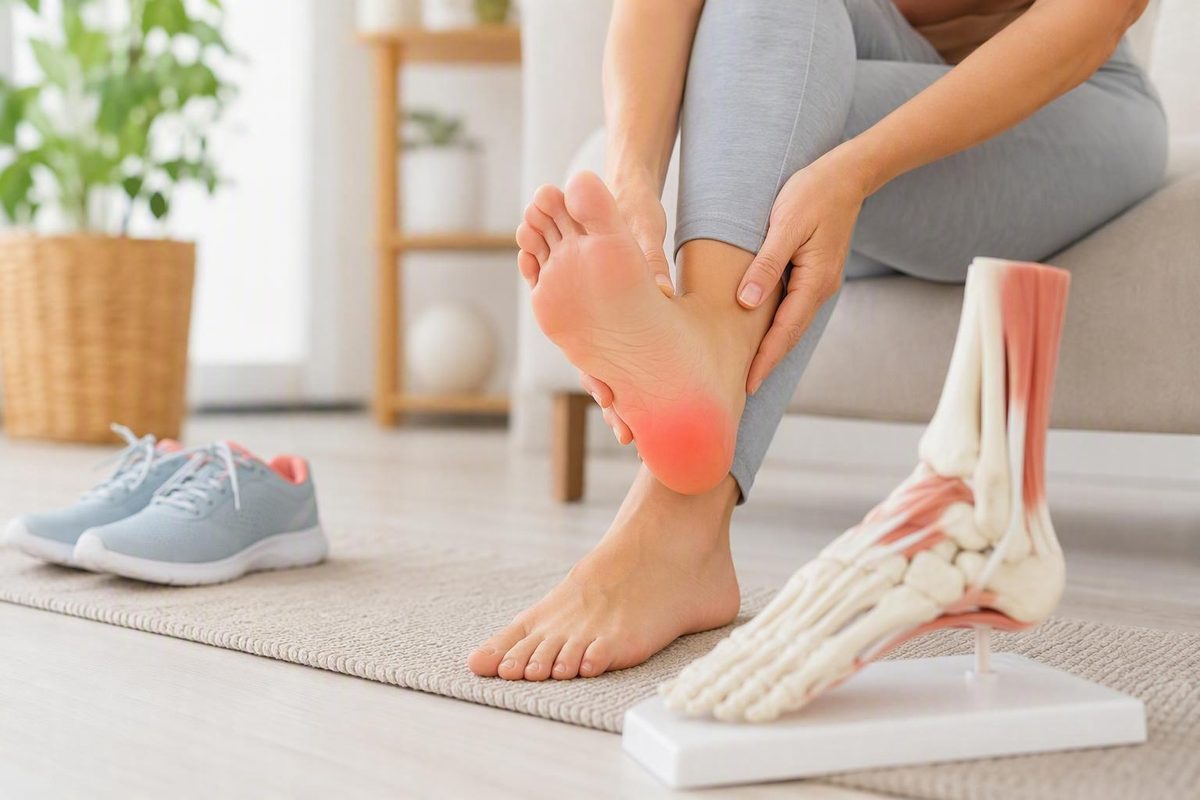
The short answer: plantar fasciitis is painful tissue; a heel spur is a bone finding
Plantar fasciitis is a disorder affecting the plantar fascia—the strong band of tissue running along the bottom of the foot—whereas a heel spur is extra bone growth projecting from the heel bone.
The practical distinction is that plantar fasciitis is commonly a source of heel pain, while most heel spurs cause no symptoms. A spur may be discovered incidentally when an X-ray is taken for heel pain or another reason. The two conditions can occur together, but plantar fasciitis can exist without a spur, and a spur can exist without painful plantar fasciitis. The American Academy of Orthopaedic Surgeons’ guidance on plantar fasciitis and bone spurs explains that most people with heel spurs do not have heel pain and that a spur generally does not need to be removed to treat plantar-fasciitis pain.
That makes the central question less “Is there a spur?” and more “Which structure is producing the symptoms?” An X-ray can confirm that extra bone is present, but the image cannot prove that the growth is causing the pain.
| Point of comparison | Plantar fasciitis | Plantar heel spur |
|---|---|---|
| Affected structure | The plantar fascia along the sole of the foot | The heel bone, usually near the plantar fascia’s attachment |
| Typical clinical clue | Pain under or near the heel with the first steps after sleep or rest | Often no clinical clue because many spurs are painless |
| Can it exist without symptoms? | The term ordinarily refers to a symptomatic disorder | Yes; a spur may be an incidental imaging finding |
| Visible on an X-ray? | Not directly shown as a routine X-ray finding | Yes |
| Usual diagnostic approach | Symptom history and physical examination, with selective imaging when needed | Identified on imaging, but symptoms and examination determine whether it is relevant |
| Likely treatment target | Painful tissue, excessive loading, flexibility, support and function | Associated painful tissue or mechanical strain—not automatically the bony growth |
| Usual role of surgery | Uncommon and considered only after prolonged nonsurgical care | Removal is rarely necessary merely because the spur is visible |
This comparison can help you understand the possibilities, but it cannot diagnose an individual case or replace medical care. The site’s general-information terms likewise characterize its foot-health articles as general reading rather than individualized medical advice. Heel pain has several potential causes, and assessment may be needed when symptoms persist, worsen or do not follow a typical pattern.
What is happening in the heel in each condition?
Despite its name, plantar fasciitis should not always be understood as a purely inflammatory problem. Depending on the case and terminology being used, clinicians may describe irritation, overload, small areas of tissue damage or degenerative change. The shared concept is that the fascia has been subjected to more stress than it is currently tolerating, producing pain near its attachment at the heel or farther into the arch. A sports-medicine review describes the condition in terms of degenerative change associated with strain and small areas of tissue damage, while acknowledging that clinical descriptions vary.
A plantar heel spur is physically different. It is a bony projection from the heel bone near the point where the plantar fascia attaches. It develops over time in association with repeated stress or tension; it does not ordinarily appear suddenly because of one routine step. Cleveland Clinic describes heel spurs as extra bone growth that develops over time in response to stress involving the foot’s ligaments or tendons.
The distinction between anatomy and symptoms matters. One person may have painful plantar-fascia tissue without a spur. Another may have a substantial-looking spur on an X-ray but no heel symptoms. When both findings are present, the painful structure may still be the plantar fascia or another nearby tissue rather than the bony projection itself.
It is also too strong to say that untreated plantar fasciitis inevitably creates a heel spur. The conditions are associated with some of the same long-term mechanical forces, particularly tension around the fascia’s attachment, but association does not establish a direct or unavoidable progression from one condition to the other.
Symptoms that suggest plantar fasciitis—and why they cannot confirm it
The characteristic plantar-fascia pattern is sharp pain under or near the heel during the first steps after getting out of bed. Similar pain may occur when standing after sitting or resting for a while. It often eases after several minutes of walking as the foot gets moving, then becomes more noticeable after prolonged standing, walking or other activity.
Tenderness commonly occurs near the underside of the heel and may extend into the arch. Some people notice pain more after activity than during it. These features are useful clues, but none is a definitive home test. Pain timing and location can overlap with other heel conditions, and the same diagnosis does not produce an identical experience in every person.
A heel spur does not have an equally dependable symptom pattern because it often produces no symptoms at all. When a person has both a spur and heel pain, the symptoms may still arise from the plantar fascia or surrounding soft tissue. Claims that a spur necessarily causes constant pain, pressure pain or pain that persists during movement are not reliable enough to distinguish the conditions: descriptions conflict, and symptoms overlap.
A practical decision aid is:
- First-step pain after sleep or rest: This raises suspicion for plantar fasciitis, particularly when the pain eases with initial movement, but it does not confirm the diagnosis.
- A painless spur found incidentally: Do not infer symptoms from the image. A structural finding does not automatically require treatment.
- Persistent, worsening or atypical pain: Arrange an assessment rather than trying to decide between plantar fasciitis and a heel spur from timing or location alone.
- Pain after an injury, numbness, substantial swelling or difficulty walking: Do not assume it is routine plantar fasciitis.
Morning pain can therefore point toward the plantar fascia, but the full picture—history, examination, activity changes and competing diagnoses—determines how much weight that clue should receive.
Why plantar fasciitis and heel spurs often appear together
Plantar fasciitis and heel spurs can coexist because both are associated with long-term mechanical stress near the plantar fascia’s attachment to the heel.
That shared setting does not mean one condition always causes the other. Longstanding tension and loading are associated with spur formation, and plantar-fascia symptoms may be present at the same time. The available evidence does not justify saying that every spur was directly caused by plantar fasciitis or that every untreated case will eventually form one.
Precise coexistence percentages are best avoided.
Consider a common clinical scenario. An X-ray shows a plantar heel spur, but pressure and loading during the examination reproduce pain along the plantar fascia. In that situation, care will generally focus on the symptomatic tissue and the forces acting on it—through activity adjustment, stretching, support and rehabilitation—rather than on eliminating the radiographic growth.
The spur may still be visible after pain subsides and normal function returns. Cleveland Clinic notes that a formed heel spur generally remains unless it is surgically removed. That does not mean treatment has failed.
How clinicians distinguish the conditions—and what imaging can and cannot show
A typical plantar fasciitis evaluation begins with a symptom history and physical examination, not routine imaging. A clinician may ask when the pain began, whether activity or footwear recently changed, what happens after rest, which activities aggravate it and whether there was an injury.
The examination may consider:
- The location where pressure reproduces tenderness.
- Whether pain is concentrated near the plantar fascia’s heel attachment or elsewhere.
- Foot and arch shape.
- Ankle movement and calf tightness.
- Gait, loading pattern and other mechanical factors.
- Recent changes in work, exercise or time spent standing.
- Footwear wear patterns and direct pressure around the heel.
- Findings that point toward another cause.
AAOS describes an examination that may assess tenderness near the heel, arch shape, limited ankle movement and signs of competing conditions such as Achilles tendinitis, heel stress fracture or nerve entrapment. Its guidance also explains that X-rays can help identify a spur, fracture, arthritis or another bony problem, while MRI and ultrasound are not routinely required for a typical plantar-fasciitis presentation.
An X-ray therefore answers a structural question: is a bony growth or other visible bone abnormality present? It cannot establish that a visible spur is the source of pain. A routine X-ray also does not directly display plantar fasciitis in the way it displays a bony projection.
Ultrasound or MRI may be considered when symptoms persist, the examination is inconclusive or another diagnosis is suspected. A podiatry-practice overview similarly notes that plantar fasciitis is usually assessed from history and examination, with ultrasound or MRI reserved for selected persistent or unclear cases.
Competing explanations for heel pain include:
- Achilles tendon problems, which generally involve the back of the heel rather than only the plantar surface.
- Stress injury, particularly when symptoms follow a marked increase in impact activity or worsen with weight-bearing.
- Nerve irritation or entrapment, which may be associated with burning, tingling, numbness or radiating discomfort.
- Arthritis, which can produce joint-related pain and stiffness.
- Shoe-related pressure, especially when discomfort is tied closely to one pair or to direct rubbing.
- Other heel-pad or soft-tissue problems, which may overlap with plantar-fascia symptoms.
These are considerations for clinical assessment, not a symptom-only diagnostic algorithm. Pain location and timing may narrow the possibilities, but they cannot reliably identify the responsible structure on their own.
The shared first-line approach: reduce strain while keeping the foot supported
Initial management overlaps substantially whether a person has plantar fasciitis alone or plantar-fascia pain alongside a heel spur. Treatment usually aims to calm symptoms, reduce excessive loading and restore comfortable function—not to treat an X-ray finding in isolation.
A staged approach may include:
- Temporarily modify aggravating activity. Reduce or pause the specific activity that repeatedly provokes pain. This does not necessarily require avoiding all movement.
- Substitute lower-impact movement where appropriate. If running or jumping aggravates the heel, an activity involving less repeated impact may help maintain movement while symptoms settle.
- Use ice for short-term symptom relief. Ice is one of the conservative measures listed in the available clinical guidance, although it does not correct the underlying loading problem.
- Stretch the calf and plantar fascia. Gentle, consistent stretching is commonly included in nonsurgical management.
- Rebuild capacity gradually. Increases in standing, walking or exercise should take account of symptom response rather than follow a rigid calendar.
- Consider physical therapy. Supervised rehabilitation may be useful when calf tightness, movement limitations, weakness, gait issues or difficulty progressing independently are contributing.
Supportive footwear, heel pads, orthotics, stretching, activity modification, ice and physical therapy are all included among the conservative options described by AAOS. These measures are options rather than guaranteed cures, and the appropriate combination depends on the individual presentation.
A practical footwear review can begin with simple questions:
- Does the shoe feel supportive and comfortable during the activities that matter?
- Has the cushioning become compressed or unevenly worn?
- Is the heel held securely, or does it move excessively?
- Does the upper press directly on a tender area?
- Do symptoms consistently worsen in one pair?
A worn or uncomfortable shoe may deserve replacement or temporary avoidance, but no single shoe style is universally required. The same caution applies to heel pads, arch supports and orthotics. Some people find that a simple insert improves comfort; others need a different shape or degree of cushioning, and some do not benefit from an insert. “Custom” does not by itself prove that a device will outperform a suitable non-custom option for a particular person.
Medication may be discussed as part of conservative care, but suitability depends on health conditions, other medicines and contraindications. This article does not provide dosing instructions. A pharmacist or clinician can advise whether a medicine is appropriate for an individual.
The overall outlook is favorable, although recovery varies. AAOS reports that more than 90% of patients improve within 10 months after beginning simple nonsurgical treatment. That figure is not an individual guarantee or a deadline.
Instead of judging recovery by a fixed number of weeks, look for trends: less intense first-step pain, greater walking tolerance, fewer post-activity flare-ups and a gradual return of function.
When symptoms persist: injections, shockwave therapy, and surgery
Escalation should follow a reassessment of the diagnosis and the response to conservative care. Continuing heel pain does not automatically mean a spur needs to be removed.
Options a clinician may discuss for persistent plantar-fascia symptoms include:
- Supervised physical therapy.
- Night splints.
- Injection-based treatments.
- Extracorporeal shockwave therapy.
- Selected minimally invasive procedures.
- Surgery in uncommon, prolonged cases.
These options are not interchangeable, and none should be presented as effective for everyone.
Corticosteroid injections require individualized risk assessment because they can weaken or rupture the plantar fascia, potentially contributing to foot flattening or chronic pain. AAOS describes the evidence for shockwave therapy as mixed and says more research is needed to determine the effectiveness of platelet-rich plasma, or PRP.
The evidence supplied here also does not establish laser therapy, dry needling, PRP, custom orthotics or another procedure as universally superior. A treatment appearing on a clinic’s service list is not proof that it is the best next step for an individual patient.
Surgery is uncommon. AAOS generally reserves plantar-fascia surgery for people who have not improved after 12 months of aggressive nonsurgical treatment. Even then, the decision should account for the diagnosis, degree of disability and possible complications—not simply the persistence of a spur on imaging.
Routine spur removal is not necessary to treat plantar fasciitis. A large spur may occasionally be addressed during an operation, but it is not usually removed merely because it appears on an X-ray. Potential surgical outcomes include continuing or chronic pain and dissatisfaction, so surgery should not be framed as an easy radiographic fix.
When heel pain needs professional assessment
Arrange an assessment when heel pain lasts more than a few weeks, continues to worsen, changes the way you walk, limits normal activity or fails to improve with reasonable conservative measures.
More prompt assessment is appropriate when pain follows an injury or is severe, when swelling is substantial, when numbness or tingling is present, or when walking or bearing weight normally is difficult. These features should not automatically be attributed to routine plantar fasciitis or an incidental heel spur. A commercial foot-health comparison also advises evaluation for persistent or worsening pain, injury-related pain, limping, severe pain, swelling, numbness or interference with daily activities.
The Shoes for Me’s standing notice separately says that sudden severe pain, spreading redness or numbness deserves clinical attention, especially for a person with diabetes. That is the publisher’s general safety notice, not a diagnosis of what those signs mean.
Atypical features matter because stress injuries, nerve disorders, arthritis, Achilles tendon conditions and other problems can resemble plantar heel pain.
The action summary is straightforward:
- A typical first-step pain pattern can initially be approached with cautious activity modification, stretching, ice and supportive footwear.
- Persistent, worsening or function-limiting pain needs evaluation.
- Severe or injury-related symptoms, neurological changes, substantial swelling, difficulty bearing weight or spreading redness should not be treated on the assumption that the problem is ordinary plantar fasciitis.
The distinction to retain is that plantar fasciitis is commonly a source of heel pain, while a heel spur is often a painless structural finding. Morning first-step pain can point toward the plantar fascia, but symptoms and imaging must be interpreted in context. Most care begins by reducing aggravating load, improving support, stretching and restoring function—not by trying to remove the spur.
Frequently asked questions
Can you have plantar fasciitis without a heel spur?
Yes. Plantar fasciitis and heel spurs are related but distinct, so plantar-fascia pain can occur when no spur is visible. A clinician can often identify a typical plantar-fasciitis presentation from the symptom history and examination without needing a spur to confirm it.
The reverse is also possible: a person may have a heel spur without painful plantar fasciitis. The presence or absence of a spur does not determine whether the fascia is symptomatic.
Can a heel spur be present without causing pain?
Yes. Most heel spurs cause no symptoms and may be discovered incidentally on an X-ray. If heel pain and a spur are present together, the image alone cannot establish whether the pain comes from the plantar fascia, another nearby tissue or a different condition.
A painless incidental spur does not need to be assigned a symptom pattern or treated merely because it is visible.
Does a heel spur go away when plantar fasciitis improves?
Usually not. A formed spur commonly remains visible even after plantar-fascia pain improves and normal function returns. That persistence does not mean the painful condition is still active or that treatment has failed.
Recovery does not require the spur to shrink or disappear. More useful measures are pain, walking tolerance, activity capacity and day-to-day function.
Do you need an X-ray to diagnose plantar fasciitis?
Not usually. A typical evaluation begins with the symptom history and physical examination. An X-ray may be considered when a clinician needs to investigate a fracture, arthritis or another bony cause, or when symptoms are persistent or atypical.
An X-ray can reveal a heel spur, but it cannot prove that the spur is causing pain. Ultrasound or MRI may be considered in selected persistent or unclear cases rather than being used routinely for every presentation.
Does a heel spur usually need to be removed?
No. Treatment normally targets painful tissue, excessive loading and impaired function rather than removing an incidental spur. Many people improve even though the spur remains unchanged.
Spur-removal surgery is rarely performed, and plantar-fascia surgery is generally reserved for prolonged, disabling symptoms that have not responded to substantial nonsurgical care. A visible spur alone is not a reason for surgery.